| Paediatric Neurosurgery | |
|
|
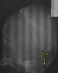
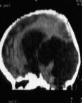
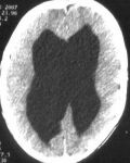
6 months Infant had onset of encephalitis science 2 weeks old, after that there is gradual swelling of his head. Brain CT scan revealed enlarge ventricules with large cyst occupying left occipital region. Fluid aspiration from the lateral venticule and cystic cavity for complete analysis to ensure there is no more infection. Surgical intervention was done with connection of both lateral ventricle and occipital cyst by Y shape connector to peritoneum cavity to decrease intracranial pressure
|
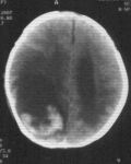
Brain CT scan revealed large cyst occupying left occipital region
Y shape connector |

Brain CT scan revealed enlarge ventricules |
|
2 days old Infant with congenital lumbosacral meningomyelocele. Operated with total excision of the sac and dural repair.
|
|

Dural patch during surgery |
|
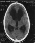
21 months old Child had recurrent vomiting with chest infection resistant to medications for months. Brain CT scan show hydrocephaly with a big cyst at the posterior fossa of her head (Dandy Walker syndrome). Operation was done with insertion of medium pressure device to the lateral ventricle connected by Y-shape connector to the posterior fossa cyst. after surgery she is very wheal and disappear vomiting.
|
Brain CT scan Hydrocephaly with a big cyst at the posterior fossa
|

Brain CT scan After surgery show shunting of both lateral ventricle and pf cyst
|
|
14 days old Child had Congenital dorsal meningomyelocele. Operated on 2-3-07 with total excision of the sac and dural repair.
|
 |
|
|
10 years old male had congenital encephalocele with hydrocephaly, ventriculoperitoneal shunt inserted since the 1st year of his life. Presented to Alumran clinic, with 4 months history of headache and repeated vomiting. During examination, he had a small mass in the back of his head with bilateral paplledema. Brain CT scan revealed hydrocephaly. Operation was done on 18-2-07 with total excision of encephalocele , insertion of new shunt and removal of the old one. During postoperative day, the patient was very well with no more headache and vomiting.
|
Brain CT scan show diluted ventricle |
Brain CT scan show encephalocele
|
| 2 days old infant had a Congenital occipital encephalocele. Operated on 10-2-07 with total excision of the sac and dural repair.
|
 |
|
|
8 years old Child with congenital hydrocephaly with ventriculoperitoneal shunting was done since the third month of his life. A mass developed in the way of his shunt for one week. Surgery was done and showed infected shunt with pus collection in the mass with consequent removal of the shunt at 16-1-07.On the first postoperative day, he developed repeated vomiting. Brain CT scan revealed intraventricular hemorrhage , urgent external ventricular shunt with heavy antibiotics cover to prevent meningitis. After 8 days, left frontal ventriculoperitoneal shunt was done. He improved gradually and discharged home very well.
|

Brain CT scan before surgery |
|
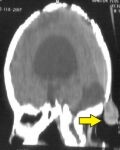
| 12 years old male presented as scalp mass at right temporal area. Brain CT scan revealed a bony erosion with soft tissue swelling. Surgical total excision with removal of unhealthy bony margin. Histopathology result showed a histiocytosis mass.
|
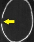
Brain CT scan bony defect |
|
|
12 years old Child had a headache and vomiting especially at morning for 14 days. On examination, she had bilateral papilledema with left facial palsy. Brain CT and MRI scanning showed dilated brain ventricles with no posterior fossa tumor. Ventriculoperitoneal shunt was done. Postoperatively she was free from any headache and vomiting and discharged home.
|
Brain CT scan diluted ventricle |
|
|
7 months old Child had repeated vomiting. Treated as gastroenteritis but not improved. Brain CT scan revealed hydrocephaly. Surgical intervention with ventriculoperitoneal shunt was done. She was very well postoperatively.
|

Brain CT scan diluted ventricle |
|
|
2 months Infant had slow deterioration of consciousness with difficulty feeding. when she reached hospital, she was unconscious and dilated right pupil with left sided paralysis. Brain CT scan showed massive right intracerebral hematoma. Urgent craniotomy was done on 16-8-06 to evacuate the hematoma. Within the 1st postoperative week, she returned to her normal breast feeding and the pupil size became normally reacting to light but still she was unable to move the right eye ball( oculomotor palsy). At the end of 1st month, she became very active child with normal movement for both eyes. |