Spine cases
 |
|
|
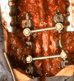
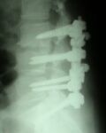
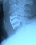
| 51 years old female had a backache more the 10 years, in the last 3 month both leg had parasthesia and numbness with frequent falling on the ground. Lumbosacral MRI show degenerative spine changes at L3-L4, central osteophyte compressed spinal roots, and a herniated disc at L5-S1. Transpedicular fixation was done with titanium roding and screw, After that L3, L4 and L5 laminectomy with excision of both L3-L4 central osteophyte and a herniated disc at L5-S1. Postoperatively the pain relieve and the patient return walking without falling in ground. |
  |
  |
| 40 years old male had ill define backache, parasthesia and numbness of both legs for one year duration. During the last one month he developed stress incontinence of urine with difficulty in defecation. The patient became unable to walk. Clinical examination show grade 3 power of hip flexors with L1 sensory level.
Dorsolumbar MRI show intradural non enhanceing small cystic nodule at the D12-L1 level compressing the spinal cord. after D12-L1 dorsal laminectomy incision to the dura was done with total excision to the mass. Histopathological result revealed cystic degeneration schwannoma. within one month postoperatively all the symptoms resolved. |
 |
 |
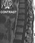
| 41 years old female presented 2 years ago complaining from difficulty to walking after an accident. In last 6 month she was became frequent falling with unable to walking. Cervical MRI show C4 -C5 disc prolapse. Anterior surgical approach to excision herniated disc and inserted prosthetic one. After that she was gradual improvement of walking. |
 |
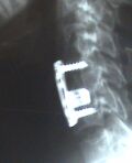
Cervical cage |
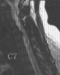
| 22 years old male had inability to stand after falling on ground for more than 5 meter height. Examination show left sided weakness more in the left leg with decrease sensation on the right. Cervical MRI show fracture in the base of fracture C5 with small fracture piece compressing the spinal cord . Anterior surgical approach to excision herniated piece, inserted prosthetic disc andC4-C6 fixation with screw and plating. After that he was gradual moving left leg. |
 |
|
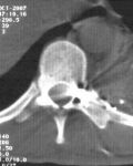
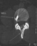
| 32 years old exposed to multiple bullet injuries to his left arm, chest and back. During examination he is unable to move left leg with loss of pain sensation in the right leg. Dorsal CT scan show bullet fragment at D10 left sided to cord while D9 bullet fragment situated anterior to the cord. During surgery D 10 laminectomy was done with removal a massive extradural lacerated muscles with shell that compressing the spinal cord to the right. After surgery he was became undergoing to physiotherapy course. After one month he was started walking without difficulties. |
  |
 |
|
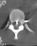
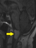
20 years old had spinal cord bullet injury perforating vertebral column with loss of his ability to walking. Brain CT scan show a fracture to L3 lamina and the bullet arrested at disc space between L3-L4. Completely removal of a fracture segment, suturing of dural tear with extraction of the bullet was done during surgery. After surgery he is gradual improvement ability for walking.
|
 the bullet arrested at disc space between L3-L4 |
 show a fracture to L3 lamina and the bullet arrested at disc space |
|
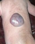
14 days old with congenital dorsal meningomyelocele. Operated on 2-3-07 with total excision of the sac and dural repair.
|
 |
|
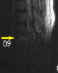
| 30 years old male exposed to road traffic accident. He had breathing difficulty with loss of the ability to move both legs. Urgent chest tube inserted to evacuate blood collection inside the chest . Dorsolumbar MRI showed severe fracture at D8-D9 with spinal cord compression. Internal vertebral column fixation with spinal cord decompression was done.
|
 |
|
|
70 years old female with a headache for one year, complete paralysis of left side of the body and weakness of the right side. Brain MRI showed anterior foramen magnum mass severely compressing the junction of brain stem and spinal cord. Total excision was done on 11-2-07, and sent for histopathology.
|
 |
 |
|
30 years old male had ataxic gait with diplopia for several years. Brain MRI showed herniated cerebellum (chiari syndrome grade 2). Surgical intervention was with craniectomy of the posterior fossa, removed C1 lamina with incised dura to be closed with patch to give more space for herniated cerebellum to be return back. After surgery, she was dramatically improved. After 3 months, ataxic gait and diplopia disappeared. Then she returned back to her work.
|
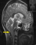 cerebellar herniation |
|
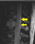
| 26 years old female case of severe backache for 4 months not responding to medication. When she visited my private clinic, she was severely ill with severe tenderness at dorsolumber region. She had rapid response to steroid. Dorsolumber MRI showed extradural hyperintese lesion extend from D11-D12. Dorsal laminectomy with complete excision of mass was done. histopathology revealed spinal cord lymphoma. she was sent for chemotherapy. Frequent postoperative MRI show complete resolution of the mass.
|
 |
 |
|
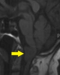
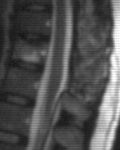
37 years old male suffered from backache and inability to walk. lumbosacral MRI showed disc prolapsed at two levels L4-L5,L5-S1.Leminectomy and discectomy was done for two levels. Post operatively, the patient started walking and after two weeks, he joined his job.
|
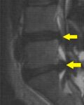 L4-L5,L5-S1 |
|