|
|
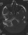
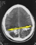
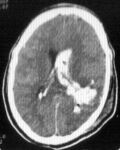
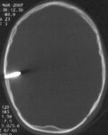
Child 10 years old had accidental bullet injury follow by a fall on ground, massive bleeding from occipital region. Brain CT scan show a bullet arrested at posterior cranial fossa. During surgery a bullet traction carefully after small incision above her cranial wound and muscle separations. Postoperatively the patient very well so discharge to home.
|

Brain CT scan show a bullet arrested at posterior cranial fossa
|
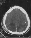
 Brain CT scan (bone window) show a bullet arrested at posterior cranial fossa Brain CT scan (bone window) show a bullet arrested at posterior cranial fossa |
|
30 years old female had fall on ground more than 5 meter height, follow by sudden loss of consciousness. Brain CT scan show large collection right parieto-occipital extradural hematoma with overlaying linear fracture extended to the base of skull. Surgery was done immediately to evacuate the extradural hematoma after craniotomy flap. Postoperatively the patient return awake without any neurological deficit.
|
Brain CT scan show large collection right parieto-occipital extradural hematoma
Brain CT scan show linear fracture extended to the base of skull |

Brain CT scan show right parieto-occipital linear fracture
|
|
21 years old male exposed to cranial bullet injury perforating his skull from left side. Brain CT scan show sever depress fracture with a bullet arrested at left temporal lobe, which have centre of speech. Completely removed of a depress segment, evacuation of hematoma, extraction of the bullet with dural patching was done during surgery. After surgery he was talking and walking normally.
|

Brain CT scan show depress fracture with a bullet arrested at left temporal lobe
|

Brain CT scan (bone window) show a bullet arrested at left temporal lobe
|
|
19 years old male exposed to perforated cranial bullet injury for his skull from left side and exit from right side with loss of his consciousness. Brain CT scan show a tract of hematoma extend from entrance to exit sit. Completely removal of a depress segment, evacuation of hematoma with dural patching was done during surgery. After surgery he was gradual improvement within ten days and return to his consciousness .
|
Brain CT scan a tract of hematoma extend from entrance to exit sit
|
Brain CT scan after two week show disappear of a tract of hematoma
|
|
33 years old male presented as a case of road traffic accident with completely loss of his consciousness. Brain CT scan show a depress segments at left frontal region with involving left orbit. Completely removal of a depress segment, reconstruction of the orbital margin with dural patching was done during surgery. After surgery he is gradual improvement and completely return his consciousness.
|

Brain CT scan fracture involving left orbit
|
Brain CT scan a depress segments at left frontal region
|
|
25 years old male had cranial shell injury with completely loss of his consciousness. Brain CT scan show a depress segments seated at left temporal region with extensive intracerebral and intraventricular hemorrhage. Completely removal of a depress segment, evacuation of hematoma with dural patching was done during surgery. After surgery he is gradual improvement completely return to consciousness.
|
Brain CT scan a depress segments with intracerebral hemorrhage at left temporal region
Brain CT scan at 7 postoperative day show disappears of depress segments, intracerebral, intraventricular hemorrhage
|
Brain CT scan extensive intraventricular hemorrhage at left temporal region
|
|
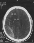
63 years old male had a headache, inability to walking and lastly loss of consciousness for one weak. He has history of mild head trauma before one month. Brain MRI show a big left sided subdural hematoma. A burr hole was done for evacuation of hematoma. Immediately after surgery the patient return oriented and he became able to walk.
|
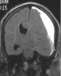
Brain MRI show a big left sided subdural hematoma
|
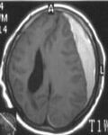
Brain MRI show a big left sided subdural hematoma
|
| Child 10 years old had Head trauma with large screw driver penetrating his skull. Brain CT scan revealed the screw driver penetrated about one inch inside his brain. Urgent surgical intervention was done to remove the screw drivercarefully under general anesthesia with repair of the brain damage. In the first postoperative day, he was very well with no serious complications.
|
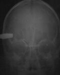
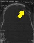
skull x-ray
|
Brain CT scan screw driverpenetrated brain
|
| 30 years old male exposed to road traffic accident. He had breathing difficulty with loss of the ability to move both legs. Urgent chest tube inserted to evacuate blood collection inside the chest . Dorsolumbar MRI showedsevere fracture at D8-D9 with spinal cord compression. Internal vertebral column fixation with spinal cord decompression was done.
|
dorsal x-ray showed fracture at D8-D9 |

MRI showed fracture at D8-D9
|
|
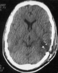
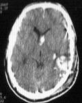
49 years old male had multiple intracranial shell injury. He was confused with impairment of vision. Brain CT scan showed two inlet at left occipital area, one of them impacted in the bone and the other perforated and arrested at the right parietal lobe. Immediate surgery was done: 1-Removal of left shell. 2-Craniectomy at left occipital bone with dural patch (site of shell entrance). 3-Craniotomy to the right parietal bone for evacuation of intracerebral hematoma. After 3 days, he got good improvement in his vision.
|
Brain CT scan 1st entrance site
|
Brain CT scan 2nd entrance site
|