| Brain Tumors | |
|
|
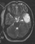
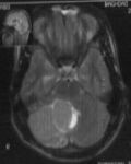
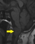
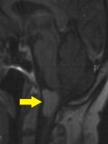
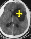
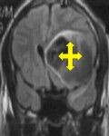
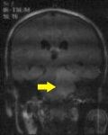
35 years old female had lethargies with enlargement of nose, lips, hands and feet. she has history of 4 years absent of menses with headache. Brain MRI show diffuse enlargement of pituitary glands with suprasellar extension. Growth hormone is so high (19.7 ng/ml). surgical intervention was done with subfrontal approach to excision of tumor. After surgery she return back to normal activity.
|
Brain MRI (coronary section) show pituitary adenoma
|

Brain MRI (sagital section) show pituitary adenoma
|
|
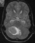
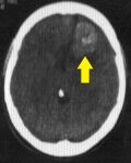
53 years old female had sudden onset of tonic-clonic fit follow by loss of her consciousness. Brain CT scan with MRI show a mass occupying left temporal lobe. Total removal with inferior temporal approach, left temporal lobe have speech centre, during surgery and send the mass for histopathology (low grade glioma). After surgery she has talking and moving normally.
|

Brain CT scan a mass occupying left temporal lobe
Brain MRI (coronal view) a mass occupying left temporal lobe |
 Brain MRI (axial view) Brain MRI (axial view)
a mass occupying left temporal lobe |
|
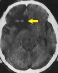
14 years old male exposed to road traffic accident with avulsed scalp before 5 year. Before 3 month he complain from a painful swelling at right sided of his head with no response to analgesia. Brain CT scan show soft tissue swelling with bone erosion at right temporal region. Completely excision of muscle and bone eroded with its margin was done during surgery and send for histopathology. After surgery the pain completely relieve. Histopathology result showed a histiocytosis mass.
|
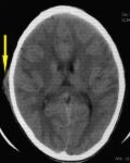
Brain CT scan soft tissue swelling at right temporal region
|

Brain CT scan bone erosion at right temporal region
|
|
young patient 19 years old had a headache with right side weakness. Brain MRI showed right cerebellar mass (PFT). Posterior fossa approach with complete excision of the mass done. Histopathologycal results show medulloblastoma, he refused radiation. The patient completely freeing from any symptom for one year, after that he became complain from repeated vomiting and right side weakness. Brain MRI showed recurrent tumour at the posterior fossa. Urgent ventriculoperitoneal shunt inserted was done to decrease intracranial pressure. After 2 weeks re-exploration and complete excision was done. The histopathologycal results show medulloblastoma nodylar type. |
Brain MRI showed PFT Brain MRI 6-3-07
showed recurrent PFT
|
Brain MRI postop. PFT disappear
|
|
35 years old female had a Headache with visual deterioration for 3 months. Repeated vomiting and inability to walk for one month. Brain CT scan revealed a 7×4 cm mass at left frontal region. Surgical intervention on 13-2-07 with very vascular tumor and she received 9 pints of blood. The mass removed completely and sent for histopathology.
|
Brain CT scan without contrast |

Brain CT scan with contrast |
| 70 years old female had a Headache for one year, complete paralysis of left side of the body and weakness of the right side. Brain MRI showed anterior foramen magnum mass severely compressing the junction of brain stem and spinal cord. Total excision was done on 11-2-07, and sent for histopathology.
|
Brain MRI without contrast |
Brain MRI with contrast |
| 65 years old male had Right sided weakness for one month with headache. Brain CT scan revealed a big right frontal mass. Operated on 4-2-07, total left frontal lobectomy was done and sent for histopathology. The result showed diffuse astrocytoma grade 2.
|
Brain CT scan |

Brain MRI with contrast |
| 17 years old male had a headache with repeated vomiting for 3 months. Brain CT scan showed a posterior fossa mass compressing the fourth ventricle causing hydrocephaly. Urgent ventriculoperitoneal shunt was done. The vomiting and headache disappeared. After 2 weeks, total removal of posterior fossa mass. The histopathology result revealed desmoplastic medulloblastoma. | 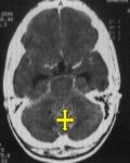
Brain CT scan before surgery |
 Brain CT scan after surgery Brain CT scan after surgery |
|
25 years old male had complaining from generalized tonic clonic attack of fit. He had history of meningitis in the first year of his life. In last 6 months, he received medication for submandibular tuberculosis. Brain MRI showed left frontal suprasylvien mass. Subtotal excision was done, postoperatively the patient became very well. Histopathology revealed diffuse astrocytoma grade 2 ,so he was sent for radiotherapy to complete his management.
|
 Brain CT scan Brain CT scan |
 Brain MRI T1 Brain MRI T1 |
|
12 years old male presented as scalp mass at right temporal area. Brain CT scan revealed a bony erosion with soft tissue swelling. Surgical total excision with removal of unhealthy bony margin. Histopathology result showed a histiocytosis mass. |
 Brain CT scan bony defect Brain CT scan bony defect |
|
|
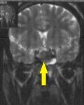
45 years old female had a headache for 2 years. In the last 6 months she developed right sided parasthesia and numbness with visual deterioration on the right eye. Brain MRI showed parasellar mass engulfing the right optic nerve and optic chiasm with pressure on the left optic nerve. Surgical intervention under microscopical field was done on 18-12-06. Complete freeing of the right optic nerve, right carotid artery and left optic nerve. She was very well in the post operative period and her vision improved gradually. Histopathological result showed meningioma.
|
 Brain MRI tumourengulf right optic nerve and carotid artery Brain MRI tumourengulf right optic nerve and carotid artery |
 Brain CT scan after surgery disappear tumour Brain CT scan after surgery disappear tumour
|
|
70 years old female had one year right sided weakness. Treated as cerebrovascular accident. In last one month she became Unable to speek. Brain CT scan showed left convexity meningioma. Surgery was done at 29-10-06 with complete excision with dural base, dural graft use to close the dural defect. In the 1st postoperative day she started to have normal speech. In the 7th postoperative day, she was discharged home with normal gait.
|
 Brain CT scan Brain CT scan
left convexity meningioma
|
|
| 33 years old male had 4 months headache with vision deterioration, and hearing loss in the left ear. On examination, bilateral papilledema with left sided facial palsy and absent left corneal reflex . Brain CT and MRI scan showed cerebellopontine angle tumor. Surgery was done with left sub occipital approach and debulking was done. On the 1st postoperative day ,the patient was very well with no more headache and gradual improvement of his vision. Histopathology result revealed schwannoma (a benign tumor with a very response to gamma knife) ,so he was sent for gamma knife for removal of ruminant part of the tumor. | 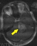 Brain CT scan Brain CT scan |
 Brain MRI Brain MRI |
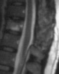
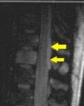
| 26 years old female had severe backache for 4 months not responding to medication. When she visited my private clinic, she was severely ill with severe tenderness at dorsolumber region. She had rapid response to steroid. Dorsolumber MRI showed extradural hyperintese lesion extend from D11-D12. Dorsal laminectomy with complete excision of mass was done on 15-5-06 histopathology revealed spinal cord lymphoma. she was sent for chemotherapy. Frequent postoperative MRI show complete resolution of the mass.
|
MRI extradural hyperintese lesion extend from D11-D12
|
11-1-07 MRI complete resolution |